Every hour you are awake, your brain accumulates metabolic debris. By the time you fall asleep, the cleanup crew has been waiting for hours.
The glymphatic system is the brain's dedicated waste clearance network. It operates almost exclusively during deep sleep, flushing toxic metabolic byproducts through channels around blood vessels using cerebrospinal fluid as the transport medium. Disrupting this process, even for a single night, measurably elevates proteins associated with neurodegeneration. Sleep is not rest for the brain. It is when the brain maintains itself.
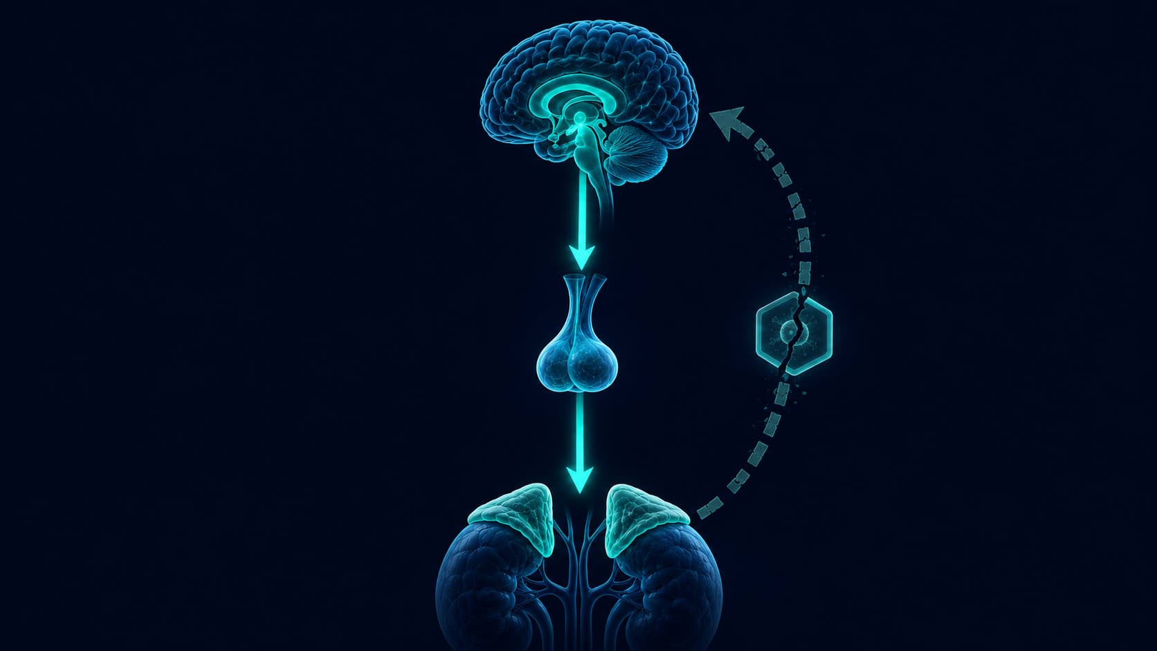
The brain is the most metabolically expensive organ in the body. It accounts for roughly two percent of body weight and consumes around twenty percent of total energy at rest. That level of activity generates waste. Proteins misfold, cellular byproducts accumulate, and the molecular environment around neurons grows progressively cluttered throughout the day. For most of the twentieth century, neuroscience had no clear answer for how any of this was removed.
Most organs rely on lymphatic vessels for this kind of maintenance. The brain sits behind the blood-brain barrier, separated from the body's general circulation, and no distinct lymphatic vessels had been identified inside the skull. The assumption, unexamined for decades, was that the brain handled waste differently. It turned out the assumption was correct. The mechanism, when researchers finally found it, was something no one had been looking for.
The System No One Had Found
A neuroscience team at the University of Rochester developed a novel application of two-photon microscopy to trace the movement of cerebrospinal fluid through living brain tissue, mapping the routes and driving mechanisms of fluid transport through the brain parenchyma in live animals.
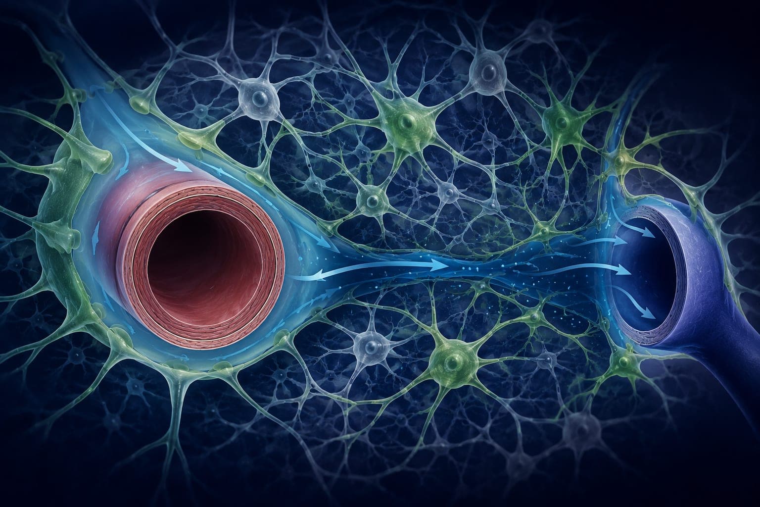
“A network of paravascular channels surrounding the brain vasculature facilitates rapid distribution of cerebrospinal fluid throughout the brain parenchyma via bulk flow, supporting clearance of interstitial proteins including amyloid-beta through a pathway dependent on astrocytic aquaporin-4 water channels.”
The pathway Iliff and colleagues described uses channels running alongside the brain's blood vessels, lined by glial cells called astrocytes. Cerebrospinal fluid enters through spaces around arteries, flows through astrocyte-lined channels, mixes with the interstitial fluid surrounding neurons, and exits through spaces around veins, carrying dissolved waste with it. The astrocytes regulate water movement through specialized aquaporin-4 channels, acting as pumps that drive the flow.
The name the team gave it, glymphatic, combines glial cell and lymphatic. The finding inverted a long-held assumption. The brain did have a dedicated waste clearance system. It was organized differently from the rest of the body, and it depended almost entirely on a state that only occurs during sleep.
What the Brain Cannot Afford to Keep
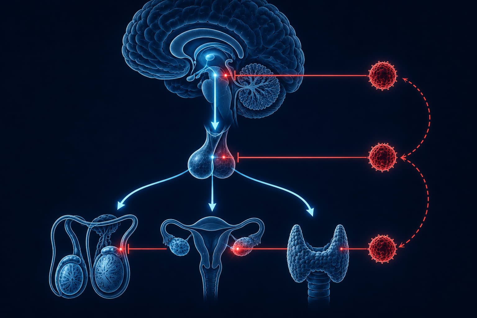
Among the waste products the glymphatic system clears most consistently are amyloid beta and tau. Both are normal proteins present in every healthy brain. Both become dangerous when they accumulate and misfold into aggregates. Amyloid beta forms plaques between neurons; tau forms tangles inside them. Both are central features of Alzheimer's disease pathology, though the precise relationship between protein accumulation and clinical symptoms involves many additional variables and remains an active area of investigation.
Amyloid beta is not a pathological abnormality. Every healthy brain produces it as a normal byproduct of neural activity. Whether it clears overnight or accumulates over decades may be, at least in part, a question of glymphatic function during sleep.
This reframes the question considerably. Alzheimer's disease is not caused by the brain producing a foreign substance. It is associated with the brain failing to clear a substance it generates every day. The research question the glymphatic discovery opened is whether some of what has been attributed to genetics or age also involves decades of suboptimal overnight clearance accumulating quietly, below any threshold of symptoms.
What Sleep Does to the Brain's Architecture
The glymphatic system does not run at a fixed rate. It requires specific physiological conditions to function at full capacity, and those conditions emerge almost exclusively during deep sleep. The same research group that identified the pathway designed a follow-up study to measure what happens to the physical dimensions of the brain's interstitial space during the transition from wakefulness to sleep, using in vivo imaging to track changes in fluid volume in living tissue.
“The volume fraction of the interstitial space increased by approximately 60% in the sleeping brain compared with the awake state, accompanied by a significantly increased convective exchange between cerebrospinal fluid and interstitial fluid and accelerated clearance of amyloid beta during sleep.”
A sixty-percent expansion in the space available for fluid movement is not a subtle shift. The brain physically reorganizes its own architecture as it enters sleep. The mechanism involves norepinephrine: during wakefulness, norepinephrine maintains astrocyte volume, keeping the interstitial space compressed. As norepinephrine falls during sleep onset, astrocytes contract, the channels widen, and the glymphatic system reaches functional capacity.
Sleep quality is therefore not a secondary variable. It is structural. Fragmented sleep, or sleep that does not sustain slow-wave stages, does not produce the same norepinephrine suppression. The channels do not open fully. Clearance runs at a fraction of capacity, and the waste that could have been removed is not.
The Waves That Drive the Clearance
Understanding how cerebrospinal fluid actually moves through brain tissue during sleep required recording three physiological signals simultaneously in humans. A research team at New York University designed a protocol combining EEG, fMRI, and flow-sensitive MRI to capture neural electrical activity, cerebral blood flow dynamics, and cerebrospinal fluid movement in parallel during overnight sleep.
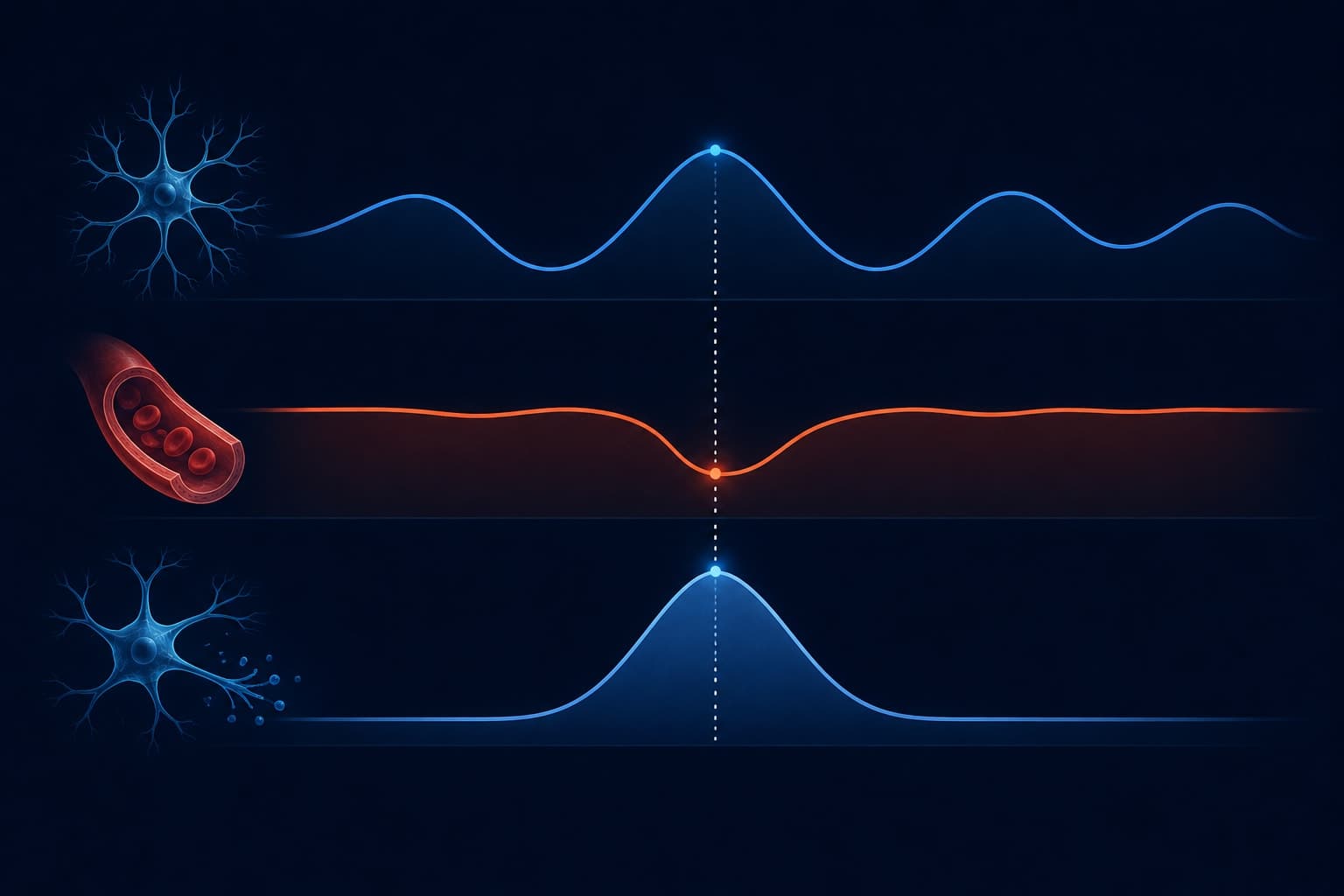
“Large oscillations in CSF flow occurred during sleep coupled to slow neural oscillations and hemodynamic changes. Each slow wave in neural electrical activity was followed by a decrease in cerebral blood volume, which preceded a large influx of CSF, suggesting that slow-wave neural activity drives pulsatile CSF movement through hemodynamic coupling.”
Each slow-wave oscillation during deep sleep creates a brief reduction in cerebral blood volume, generating a pressure gradient that pulls cerebrospinal fluid into the brain in a distinct pulse. These are not gradual trickles. The flows Fultz and colleagues recorded were large, rhythmic, and tightly synchronized with the electrical architecture of slow-wave sleep.
More slow-wave sleep produces more pulses. Fewer pulses mean less fluid transported through tissue. Less fluid movement means less waste carried out. The glymphatic system is not a continuous background process. It is event-driven, and the events are deep sleep oscillations.
What Gets Left Behind
The most direct human evidence for what glymphatic disruption produces comes from controlled sampling of cerebrospinal fluid composition. A research group at Washington University designed a within-subject study to measure tau protein concentrations in human cerebrospinal fluid following a normal sleep night and following a single night of acute sleep deprivation, using paired samples drawn under both conditions from the same participants.
“Acute sleep deprivation produced a significant increase in tau concentration in human cerebrospinal fluid compared with the normal sleep condition, consistent with impaired interstitial clearance during extended wakefulness.”
A measurable shift in protein composition after a single disrupted night reframes what chronic sleep restriction actually accumulates. The proteins do not wait for months of impairment to begin rising. The deficit is immediate.
One disrupted night does not cause neurodegeneration. The relationship between short-term tau elevation and long-term clinical outcomes involves many additional factors, and this area of research is still developing. What the data supports is that the clearance deficit is real, begins within hours, and repeats across every disrupted night for as long as the pattern continues.
Why the System Deteriorates With Age
Glymphatic efficiency is not fixed across the lifespan. A research team examined glymphatic pathway structure and function across the rodent lifespan, measuring both the anatomical properties of paravascular channels and cerebrospinal fluid flow rates in young adult, middle-aged, and aged animals.
“Glymphatic pathway activity was significantly reduced in aged animals compared with young adults, associated with a loss of polarized aquaporin-4 localization at astrocyte endfeet lining the paravascular channels.”
The mechanism behind the age-related decline centers on aquaporin-4 polarity. In younger brains, these water channels are concentrated at the astrocyte endfoot surfaces facing blood vessels, precisely where they need to be to drive fluid movement efficiently. In aging tissue, that distribution becomes disrupted. The pump still exists, but it progressively loses mechanical advantage at the cellular level.
Two separate failures converging on the same outcome. Older adults tend to spend less time in slow-wave stages, which reduces the frequency of the CSF pulses Fultz and colleagues mapped. Channel efficiency simultaneously declines. Less pumping capacity compounds less pulse frequency: less waste cleared per night, every night, accumulating across years.
Optimized clearance
- Abundant slow-wave sleep generating frequent CSF pulses
- Aquaporin-4 channels polarized at astrocyte endfeet
- Interstitial space expands fully during sleep
- Amyloid beta and tau cleared toward lymphatic drainage
- Consistent sleep timing preserves early-night slow-wave window
Impaired clearance
- Fragmented or shortened sleep reduces CSF pulse frequency
- Aquaporin-4 polarity disrupted with aging and chronic poor sleep
- Partial channel opening limits fluid throughput
- Protein accumulation rate exceeds clearance rate
- Late or irregular sleep timing compresses the slow-wave window
What the Research Actually Changes
Several findings in this field, particularly around exact clearance rates and their direct relationship to disease outcomes, remain more provisional than others. The research base is still being extended from animal models to human physiology. What the evidence supports consistently is the relationship between slow-wave sleep, cerebrospinal fluid dynamics, and the removal of metabolic waste. Three variables are most directly within reach.
Slow-Wave Architecture Matters More Than Duration
The glymphatic system's clearance capacity tracks slow-wave activity, not simply hours in bed. Alcohol consumed within four hours of sleep, benzodiazepines, and most antihistamine sleep aids suppress slow-wave architecture while often maintaining total sleep duration. An alcohol-assisted eight hours may provide substantially less glymphatic clearance than six hours of consolidated, undisturbed deep sleep. Duration is a floor condition. Slow-wave activity is the active variable.
Critical mechanismSleep Timing Compresses the Slow-Wave Window
Slow-wave sleep is concentrated in the first half of the night, in alignment with the circadian system. Sleeping significantly later than your habitual schedule compresses this window even when total sleep duration is preserved. An eight-hour sleep beginning at 2am contains substantially less slow-wave activity than seven hours beginning at 11pm. Glymphatic clearance is not distributed evenly across a night's sleep.
Often underestimatedCore Temperature at Sleep Onset
Core temperature reduction at sleep onset is associated with the norepinephrine drop that allows glymphatic channels to open. A cool sleeping environment supports this. A warm bath or shower taken 90 minutes before bed accelerates the process through peripheral vasodilation, drawing heat from the core and facilitating the temperature drop. The direct research on glymphatic function and temperature is still developing, but the sleep-onset mechanism is well established.
Emerging evidence- Brief naps: insufficient duration for sustained slow-wave clearance activity
- Stimulants: manage waking performance but do not address accumulated waste
- Weekend recovery sleep: may partially restore clearance but cannot fully reverse a chronic deficit
- Supplements marketed as brain detox products: no established evidence of glymphatic augmentation
- Fragmented or light sleep: maintains some rest function but leaves clearance channels only partially open
The brain is not a passive recipient of sleep's benefits. Every night of consolidated deep sleep is, among other things, a hydraulic operation: channels opening, fluid moving, proteins transported from the interstitial space toward the brain's vascular margins and from there to the body's clearance systems. The process evolved over hundreds of millions of years and runs on a schedule the brain cannot renegotiate.
Sleep is not when the brain rests. It is when the brain maintains itself.
Science-backed insights, twice a week
The GetClariSync Body Desk publishes research summaries on neuroscience, biological aging, and the mechanisms behind how the body maintains itself. No opinion pieces, no self-help clichés.
Join the newsletterGetClariSync Body Desk
Editorial Research · Sports & Movement Science
The GetClariSync Body Desk reviews research in exercise physiology, recovery science, and sports nutrition. We follow journals including Medicine & Science in Sports & Exercise, the Journal of Applied Physiology, the British Journal of Sports Medicine, and the European Journal of Applied Physiology. We separate findings from trained-athlete populations from those relevant to recreational readers, and we flag when transferring a protocol across populations is unsupported. We are editorial researchers, not certified trainers, physiotherapists, or sports physicians — please consult a qualified professional before starting new exercise programs, especially with existing injuries, pregnancy, cardiovascular conditions, or chronic disease.